A sample preparation procedure enables acquisition of 2-channel super-resolution 3D STED image of an entire oocyte
Michaela Frolikova, Michaela Blazikova, Martin Capek, Helena Chmelova, Jan Valecka, Veronika Kolackova, Eliska Valaskova, Martin Gregor, Katerina Komrskova, Ondrej Horvath, Ivan Novotny
bioRxiv 2023.03.07.531472; doi: https://doi.org/10.1101/2023.03.07.531472
ABSTRACT
Super-resolution (SR) microscopy is a cutting-edge method that can provide detailed structural information with high resolution. However, the thickness of the specimen has been a major limitation for SR methods, and larger structures have posed a challenge. To overcome this, the key step is to optimize sample preparation to ensure optical homogeneity and clarity, which can enhance the capabilities of SR methods for the acquisition of thicker structures.
Oocytes are the largest cells in the mammalian body and are crucial objects in reproductive biology. They are especially useful for studying membrane proteins. However, oocytes are extremely fragile and sensitive to mechanical manipulation and osmotic shocks, making sample preparation a critical and challenging step.
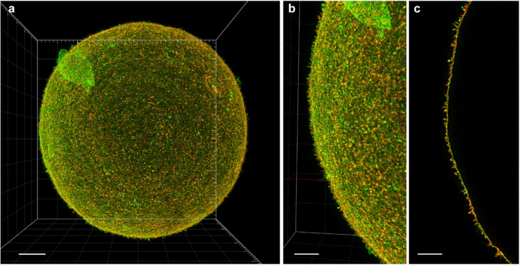
We present an innovative, simple, and sensitive approach to oocyte sample preparation for 3D STED acquisition. This involves alcohol dehydration and mounting into a high refractive index medium. This extended preparation procedure allowed us to successfully obtain a unique 2-channel 3D STED super-resolution image of an entire mouse oocyte.
By optimizing sample preparation, we can overcome the limitations of SR methods and obtain high-resolution images of larger structures, such as oocytes, Knowledge of which are important for understanding fundamental biological processes.
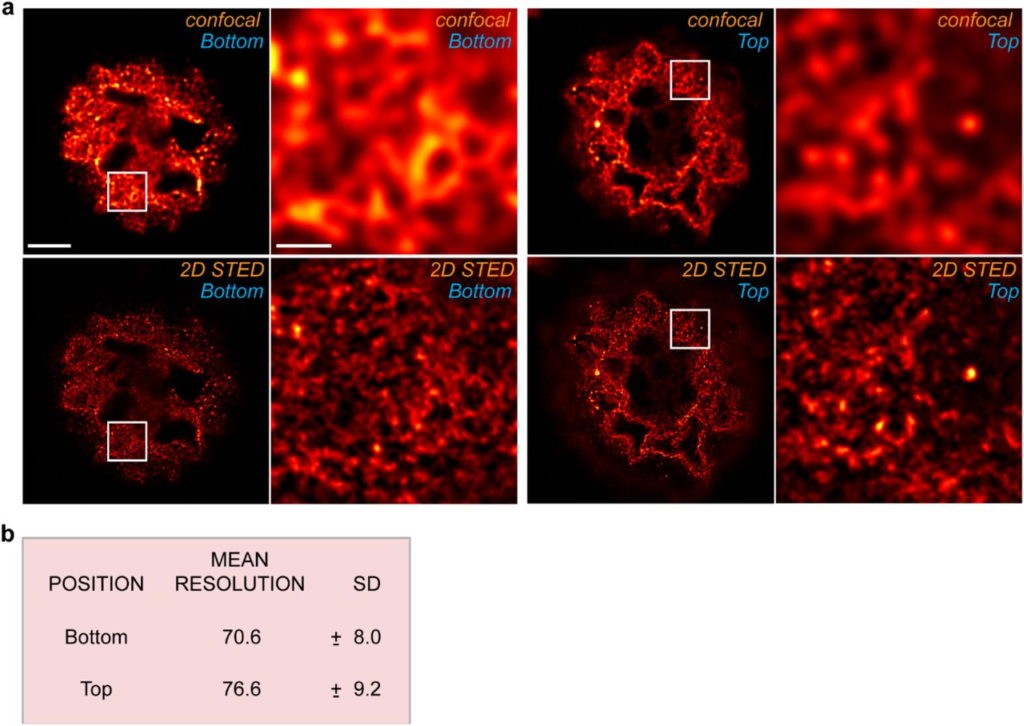
Achieved resolution: